Product description
Product Description
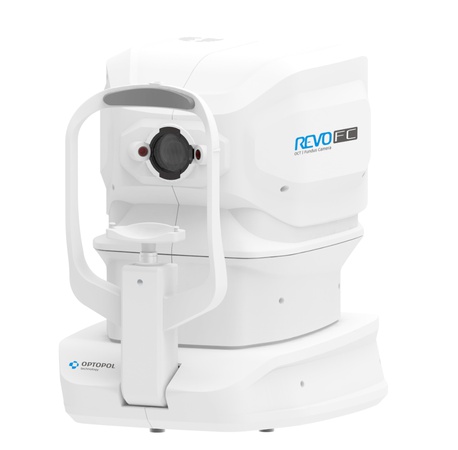
The built-in 12.3 Mpix camera guarantees excellent image reproduction. REVO FC meets all requirements for modern optical tomographs.Fundus Camera with the complete REVO 80 functionalityThe combination of an All in One OCT technology with a Full Color Fundus Camera in one compact system gives you high quality OCT images and a detailed color image for a multipurpose diagnosis. Capturing color fundus images of retina and OCT scanning of a retina in a single shot on one device saves both time and space.Now you can use the REVO FC in the way you need it:- As a Full Color Fundus Camera- As a combo providing simultaneous OCT and fundus images- Only for high quality OCT imaging including OCT-A- As a Biometry deviceREVO FC offers all proven advantages of REVO systems with a cutting-edge color Fundus imaging for a new level of diagnostic certainty. High quality OCT scanning and a comprehensive analysis of the retinal layers combined with a Fundus imaging make the examination versatile as never before.What makes the REVO FC truly unique is its integrated non-mydriatic 12.3 Mpix Fundus Camera capable of capturing ultra-high quality and detailed color images. The REVO FC Fundus Camera is fully automated, safe and easy to use.- The advanced optical system ensures high quality imaging at as wide as 45° viewing angle.- A Color Fundus image capturing is possible with a pupil as small as 3.3 mm. An OCT scan is possible even on a 2.4 mm pupil.- Easy to use image processing tools such as RGB channel, brightness, contrast, gamma and sharpness adjusters used with filters deliver a stunning retinal image. - Available view modes present detailed photos of a single or both eyes as well as a time comparison of the fundus photos.RETINASingle 3D Retina examination is enough to perform both Retina and Glaucoma analysis based on retinal scans. Software automatically recognizes 8 retina layers. Thus allowing a more precise diagnosis and mapping of any changes in the patient’s retina condition.GLAUCOMAComprehensive glaucoma analytical tools for quantification of the Nerve Fiber Layer, Ganglion layer and Optic Head with DDLS allow for the precise diagnosis and monitoring of glaucoma over time.With the golden standard 14 optic nerve parameters and a new Rim to Disc and Rim Absence the description of ONH condition is quick and precise.Advanced view which provides combined information from Retina and Disc scan to integrate details of the Ganglion cells, RNFL, ONH in a wide field perspective for comprehensive analysis. Specifications:- Type: Non-mydriatic fundus camera- Photography type: Color- Angle of view: 45° ± 5%- Min. pupil size for fundus: 3.3 mm or more- Camera: Builit-in 12-megapixel CCD camera- Light Source: SLED, Wavelength 830 nm- Bandwidth: 50 nm half bandwidth- Scanning speed: 80 000 measurements per second- Axial resolution: 2.6 μm digital, 5 μm in tissue- Transverse Resolution: 12 μm, typical 18 μm- Overall scan depth: 2.4 mm- Focus adjustment range: -25 D to +25 D- Scan range: Posterior 5–12 mm, Angio 3–9 mm, Anterior 3–16 mm- Scan types: 3D, Angio*, Radial (HD), B-scan (HD), Raster (HD), Cross (HD), TOPO, AL, ACD- Fundus alignment: IR, Live Fundus Reconstruction- Alignment method: Fully automatic, Automatic, Semi Manual- Retina analysis: Retina thickness, Inner Retinal thickness, Outer Retinal thickness, RNFL+GCL+IPL thickness, GCL+IPL thickness, RNFL thickness, RPE deformation, IS/OS thickness- Angiography OCT: Superficial plexus, RPCP, Deep Plexus, Outer Retina, Choriocapilaries, Depth Coded, Custom, Enface, FAZ, VFA, NFA, Quantification: Vessel Area Density, Skeleton Area Density, Thickness map- Glaucoma analysis: RNFL, ONH morphology, DDLS, OU and Hemisphere asymmetry, Ganglion analysis as RNFL+GCL+IP and GCL+IPL- Angiography mosaic: Acquistion method: Auto, Manual- Mosaic modes: 10x6 mm, Manual up to 12 images- Biometry OCT: AL, CCT, ACD, LT- Anterior Wide (No lens/adapter required): Pachymetry, Epithelium map, Angle Assessment, AIOP, AOD- 500/750, TISA 500/750, Angle to Angle view- Connectivity: DICOM Storage SCU, DICOM MWL SCU, CMDL, Networking- Fixation target: OLED display (The target shape and position can be changed), External fixation arm- Dimensions (WxDxH) / Weight: 367 x 480 x 504 mm / 30 kg- Power supply / consumption: 100-240 V, 50/60 Hz / 115-140 VA
Learn more


